
Early research results
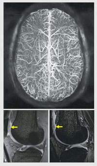 |
During the first year after the commissioning of the 7-Tesla MR imager, scientists working at the Institute have already showed that the higher structural resolution and greatly improved contrast offered by the 7 Tesla permits much better evaluation of anatomical structures including the hippocamus. The hippocamus is part of the limbic system involved in memory formation and displays characteristic changes in patients with advanced Alzheimers disease. There is thus a good perspective to counter the drastic health consequences of this disease with earlier diagnosis and commencement of therapy in the foreseeable future. In the brain it could also be shown that the venous system can be visualized with a heretofore unknown richness in detail without the use of contrast agent. Comparable images at 1.5 Tesla are not feasible; the 7-Tesla images are based on the pronounced magnetic properties of deoxygenated blood. Outside the head it could be shown for the first time that cartilage changes can be imaged with higher sensitivity and richness in detail compared to conventional 1.5 Tesla imaging. New innovative drugs are in some cases effective in slowing down or halting the progression of degenerative changes (osteoarthritis), but only when the intervention occurs at an early stage. Of particular importance for further dissemination of 7-Tesla technology: Hahn scientists were able to demonstrate in a study with more than 100 volunteers that, although transient side-effects (vertigo, metallic taste) at 7 Tesla occurred more often compared to 1.5 Tesla, the examination is well tolerated in the majority of cases. In this regard the implementation of this high field strength into clinical routine seems unproblematic.