Research
There are now eight research groups working at the Erwin L. Hahn Institute for MR Imaging. The main research interests and expertise of these groups cover very different areas of specialisation and application, which permits both complementary and synergistic collaboration. The close interdisciplinary and international cooperation between the research groups enables the ELH to investigate technical, methodological and medical questions relating to 7 Tesla UHF MRI across all the disciplines – a unique feature that helps to make the Institute one of the world’s leading centres for UHF MRI research and application. The main areas of research are divided into very different and nonetheless complementary disciplines and fields.
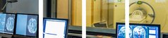
Methods & Technologies – Making UHF MRI Fit for Clinical Practice
One of the main interests of the ELH is in advancing UHF MRI. Various research groups at the ELH work on the development and research of new technologies and their methods of application with the aim of making high-resolution magnetic resonance imaging applicable in clinical and diagnostic practice.
Radio-frequency technology
Research in the group of Prof. Dr. Mark Ladd of the German Cancer Research Center (DKFZ) in Heidelberg centres on the development of methods and technologies to make 7 Tesla examination possible in all parts of the human body, including the torso. The particular focus here is on the following: radio frequency (RF) excitation antennas with several independent elements, numerical simulation in inhomogeneous models of the human body in order to explore distribution of the transmitting magnetic field (B1) and the associated temperature increase in the body (SAR), including in the presence of electrically conducting implants, and radio-frequency excitation strategies for more even distribution of the B1 field or spatially selective excitation/saturation.
In cooperation with the group of Prof. Dr. Harald H. Quick, work is also ongoing as part of a DFG-funded project (Deutsche Ultrahochfeld- Bildgebung/German Ultrahigh Field Imaging, GUFI) at various locations on quality assurance standards for MR imaging with very powerful magnetic fields.
The ELH in Essen, the DKFZ in Heidelberg and the Institute of Microwave and RF Technology in Duisburg (Prof. Dr. Klaus Solbach) are also working as part of a collaborative project on research and development of a 32-channel RF transmit system that is the only one its kind in the world to date. Up to now, 7T UHF MRI systems have had a maximum of 16 independent RF transmit channels, which means that the groups are undertaking pioneering work in this area. The project was funded until April 2017 by the European Research Council through its (ERC) Advanced Grant “MRexcite”.
Radio-frequency antennas and diagnostic Applications
The High Field and Hybrid MR Imaging research group led by Prof. Dr. Harald H. Quick develops and evaluates new technologies and methods for expanding the range of clinical applications for 7 Tesla UHF MRI. More specifically, the group simulates, develops and builds new multi-channel RF transmit/receive coils for UHF MRI imaging and signal homogenisation methods for 7T neurological and body UHF MRI. The aim is to fully exploit the high signalto- noise ratio (SNR) of UHF MRI and achieve the highest possible functional and spatial detail resolution for various applications in brain and body UHF MRI. The research groups of Prof. Harald Quick and Prof. Mark E. Ladd (DKFZ Heidelberg) work in close cooperation in this area.
Active cooperation also exists with various clinical users of 7T UHF MRI at Essen University Hospital. The advantages and disadvantages of 7 Tesla UHF MRI in relation to standard MRI at 1.5 and 3.0 Tesla are evaluated in comparative clinical studies. The research groups within the Erwin L. Hahn Institute that have a chiefly neurological focus benefit through this process from new RF head coils and methods. New RF body coils can further improve high-resolution oncological MR imaging and help to extend its application to other parts of the body (thorax, abdomen, pelvis); active cooperation in this area is currently underway with Dr. Tom Scheenen’s research group.
Another main research interest of the High Field and Hybrid MR Imaging research group is safe application of MRI for patients with passive and active implants.
Cancer diagnosis
Dr. Tom Scheenen’s research group specialises in advancing MR imaging and MR spectroscopy for oncological applications and their transfer into clinically relevant use. Research work in this group ranges from development of new RF coil technology and imaging sequences for 7 Tesla UHF MRI, through investigation of new in-vivo biomarkers to assess the aggressiveness of cancer, especially prostate cancer, to large-scale patient studies. Based on the excellent research results on prostate diagnosis, the scope for cancer diagnosis using 7 Tesla UHF MRI is to be extended in future to also include visualisation of the smallest metastases of different types of tumour.
High-resolution neuroimaging
Research in Dr. Koopmans’ group aims to improve spatial detail precision in neuroimaging (fMRI & DWI). High-resolution MRI measurements present a number of challenges for scanner performance. Compared with standard MRIs, the data points acquired in UHF MRI are 10 to 100 times closer together. A large part of Dr. Koopmans’ research is therefore devoted to accelerating the process, both on the signalexciting side (multiband RF pulses that are compatible with the ultra-high magnetic field strengths) and in signal reconstruction (parallel imaging techniques).
The second focus in the group is on a specific area of application for high-resolution fMRI: imaging individual layers of the cerebral cortex. Conventional fMRI (approx. 2–3 mm precision) cannot capture the layers individually, as they are less than 1 millimetre thick. With the support of the DFG’s Emmy-Noether Programme and as one of the pioneers in the field of layer-specific fMRI, Dr. Koopmans now aims to improve the imaging techniques and develop a layer-specific analytical tool and signal models. Pain imaging is the evidence base for his work. Here the layers of the cerebral cortex should deliver insights into how information is processed in the brain and the spinal cord.
Functional MRI – Inside the Human Head
For the research groups introduced so far, the main focus of their work is directly on 7 Tesla MRI and its technical advancement and application. The other research groups at the ELH work chiefly in functional MRI (fMRI), a technique that makes it possible to map brain activity and therefore helps to explain thought processes, for example.
Neurospectroscopy
Gamma-amino butyric acid (GABA) is the main inhibitory neurotransmitter in the brain and can be detected using MRI proton resonance spectroscopy. However, the weak signal is masked by signals from other metabolites. The research group led by Prof. Dr. David Norris has succeeded in implementing techniques for 7 Tesla MRI spectroscopy which make it possible to detect the GABA signal.
The group’s work is divided into two parts: improving measurement methods, and applications chiefly in the field of diabetes research. In relation to the measurement methods, the aim is to make experiments more robust against unwanted but unavoidable variations in the static and radiofrequency magnetic fields. The area of application is to be extended by developing new methods of measuring the relative distribution of metabolites between white and grey matter. Improving quantification by correcting the unwanted macromolecule signal on the GABA signal is another focus of the group’s work. In diabetes research, investigation is also underway on the relationship between memory performance and the GABA concentration in certain areas of the brain.
In a joint study with the German Diabetes Center (DDZ), it has been shown that diabetics have poorer memory performance than healthy subjects, and that there is a correlation between performance and GABA concentration in the medial prefrontal cortex. In another area of the brain that is important for memory, the precuneus, no correlation was found.
Decision-making and behavioural addictions Research
The research group of Prof. Dr. Matthias Brand is interested in neural correlates of cognitive and emotive processes. A particular focus of the research here is on how decision–making can be influenced by emotion processing, manmachine interaction, and the neurobiological and neuropsychological principles of behavioural addictions, such as internet addiction or pathological buying. The research primarily looks at brain responses to stimuli associated with addictions and their significance for subjectively perceived craving. The high magnetic field strength and accompanying good spatial resolution of the UHF MRI facilities at the Erwin L. Hahn Institute make it possible to achieve internal differentiation of individual brain structures, such as the amygdala or the ventral striatum. For the fMRI research outlined, the 7 Tesla MRI system also allows visualisation of activations in small structures, which is not possible or only with great difficulty in 1.5 or 3.0 Tesla MRI.
Function of the cerebellum
The high field strength of the MRI at the Erwin L. Hahn Institute is also extremely beneficial for research into the cerebellum. For example, investigation of the cerebellar nuclei located deep in the cerebellum is improved significantly by the use of 7 Tesla UHF MRI and in some cases would be impossible without it. Contrary to belief for many years, the cerebellum not only supports motor and learning processes but also plays a role as a modulator in many other areas, including certain cognitive functions, emotional processing, and pain. It is therefore attracting increasing interest in the neurosciences.
The Experimental Neurology group led by Prof. Dr. Timmann-Braun uses UHF MRI for structural visualisation of the cerebellar nuclei in healthy subjects and in patients with certain conditions affecting the cerebellum (known as ataxias), and also for functional MRI studies. As part of a Collaborative Research Centre funded by the DFG (SFB 1280 Extinction Learning; coordinator: O. Güntürkün, RUB; co-coordinator: D. Timmann-Braun), work is currently focusing on the significance of the cerebellum for the extinction of learned fear responses. Although it has been known for a long time that the cerebellum plays a role in learning associations, including anxiety conditioning, little is known about its importance to the capacity for “unlearning” (extinction). Extinction processes play a major role in anxiety disorders, and likely also in chronic pain disorders. Dr. Timmann-Braun is working with Dr. H.-H. Quick and using UHF MRI to test the hypothesis that the cerebellum plays an important role in the neuronal network involved in the extinction of learned fear.
Pain research
The research group of Prof. Dr. Ulrike Bingel uses high-resolution MR imaging of the brain stem and spinal cord to investigate the connections between certain subcortical areas and pain processing in the spinal cord. One of the main interests in this group is to explore the interface between pain processing in the central nervous system and cognitive neurosciences. The researchers are investigating the mechanisms of individual pain perception, vulnerability to chronification of pain, and the capacity to modulate pain in certain contextual circumstances. Structural and functional MRI are used here in combination with pharmacological and psychophysical methods. The studies are conducted on healthy subjects and groups of patients suffering from chronic pain or neurological conditions such as Parkinson’s disease. Current studies aim to advance understanding of interindividual differences in response to placebos in pharmacological therapies in order to further improve therapy management.