Erwin L. Hahn Institute for MRI
Research at the Erwin L. Hahn Institute covers three main research areas: methodology development and safety at 7T, clinical imaging at 7T, and functional MRI at 7T. The latter includes not only research into cognitive science topics but also spectroscopy, with which metabolites can be analysed in a non-destructive manner.
The chief advantage of ultra-high field MRI is the increased spatial resolution it offers for both anatomical imaging and functional MRI. Its main perceived drawback has been non-uniformities in the transmit/receive radiofrequency fields. These problems are being addressed by the methodology and safety research area. The research focuses on the development of RF coils, sophisticated parallel transmit approaches, and new reconstruction methods, with special attention to clinical use. Further central topics deal intensively with the safety of developed components and the influence of ultra-high field MRI on the human body. The Erwin L. Hahn Institute is in a unique position to tackle these problems, having one of the world’s leading groups specialized in mastering the effects of non-uniformities in the radiofrequency magnetic fields, particularly in cardiac and abdominal imaging. A system developed and built by the group to manipulate the transmit field was successfully patented in 2010 and now permits clinical imaging of the trunk of the body.
The strong interdisciplinary cooperation between clinicians, engineers and scientists has been instrumental in making clinical imaging of the entire human body, and particularly pathologies, at 7T a unique feature of the Erwin L. Hahn Institute. In many of its projects, anatomical regions and pathologies have been depicted for the very first time at 7T, and comparative studies with 1.5T and 3T have revealed the first promising clinical benefits of 7T. Its close ties to University Hospital Essen give the Institute access to a broad range of pathologies and the possibility of collaborating with outstanding clinical partners.
With the increasing number of clinically oriented studies at 7T, scientists face the challenge of providing new coil concepts for ultra-high field MRI in body parts other than the head. Large field-of-view imaging is important for assessing patients with metastases or multiple sclerosis lesions in the spinal cord, for example. In 2011, the first results worldwide were published for imaging of abdominal organs with contrast agents. Erwin L. Hahn Institute scientists were able to implement novel methods to improve imaging at 7T even in such challenging anatomical regions. Currently, work is underway to assess a variety of pathologies in patients to further elucidate the clinical impact of this technology.
In functional brain imaging, 7T enables activated brain areas to be resolved with high resolution. This makes it possible for the Institute’s scientists to study activated brain areas to reveal the neurobiological basis of cognitive skills and other aspects of human behaviour. Such studies are performed to obtain additional information about both patients and healthy subjects. The significantly higher spatial resolution of fMRI at 7T allows new insights into the role of even the smallest brain structures in various cognitive and emotional processes. In addition, the studies contribute to a better understanding of the neurobiological basis of various psychological symptoms in patients with brain disorders. 7T provides great opportunities for such fMRI studies, in particular due to the improved signal-to-noise ratio. Imaging sequences are also continually being optimized at the Erwin L. Hahn Institute to make the benefits of ultra-high field MRI available to functional brain imaging.
With regard to spectroscopic applications, the benefits of ultra-high magnetic field systems are two-fold: not only does the sensitivity for the detectable metabolites increase, but also the spectral resolution itself. The resulting spectra contain metabolic fingerprints of the given tissue. In the prostate, for example, this fingerprint can be used to discriminate between cancerous and non-cancerous tissue.

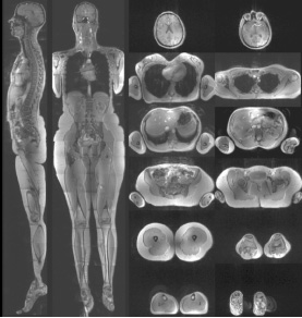
Head-to-toe images from a human volunteer acquired with a 2.08 mm isotropic resolution. The dataset was obtained with a moving table in 28 stations. The slices are fairly homogeneous even though a non-optimized combination of the CP+ and CP2+ modes was used throughout the acquisition, demonstrating the robustness of the method.

(A) T2-weighted HASTE image in a human volunteer; note the absence of signal dropouts. (B) Gradient echo image with fat saturation and (C) gradient echo image with water saturation; the saturation is effective throughout the entire slice.
